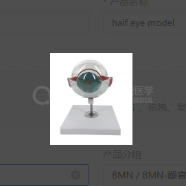
BIX-A1062
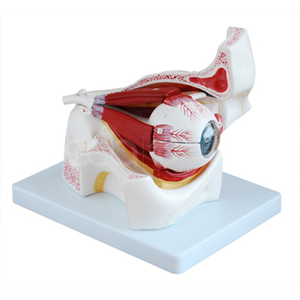
1.This model shows the position of the eye and optic nerve in the eye socket (bottom and middle wall). 2.Divided into 10 pieces. 3.times magnification.
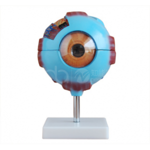
ADA-316A Giant Eye Model — White Eyeball Anatomy Teaching Model
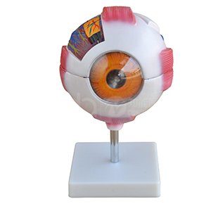
BIX-A1061 Eyeball Amplification Model 6X — Human Eye Anatomical Structure Model
BIX-A1060 Common Eye Disease Model 20pcs/Set — 3X Enlarged Eye Pathology Teaching Set
BIX-LV14 Intraocular Retinopathy Inspection Training Eye Model — 13 Fundus Lesion Modules
BMN/S035 Half Eye Model 3X Enlarged — 5-Part Midsagittal Eyeball Anatomy Model