


Half body CPR training manikin(Sim....
BIX/CPR100A
Advanced fully automatic electroni....
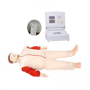
BIX/CPR480
Advanced computer half body CPR ma....
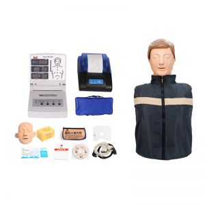
BIX/CPR260
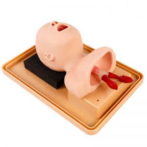
Advanced infant head for trachea i....
BIX-J3A
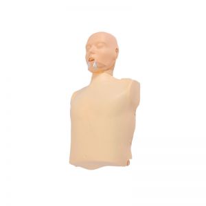
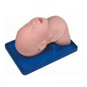
Neonate Head for trachea Intubatio....
BIX-J2A
Created on:2025-11-01 | bomn
The cross-sectional anatomical model of the human neck is a high-precision teaching tool used in medical education and clinical training, aiming to demonstrate the complex anatomical layers and spatial relationships of the neck.
...The cross-sectional anatomical model of the human neck is a high-precision teaching tool used in medical education and clinical training, aiming to demonstrate the complex anatomical layers and spatial relationships of the neck. This model precisely reproduces tissues such as skin, muscle, blood vessels, nerves, trachea, esophagus, cervical vertebrae and lymph nodes through multi-layered cross-sectional structures. It uses realistic colors to distinguish different tissue types, enabling learners to clearly understand the mutual positions and functional connections among various structures in the neck.
In teaching, the cross-sectional anatomical model of the neck helps students master the stratified anatomical features of the neck, visually demonstrating the course paths of important structures such as the carotid sheath, jugular vein, common carotid artery and vagus nerve. It is an important auxiliary material for learning courses such as head and neck surgery, imaging and anesthesiology. Students can understand the anatomical structures corresponding to imaging sections such as CT and MRI through observation and stratified disassembly, thereby enhancing their image recognition ability and spatial thinking.
In addition, this model is widely applied in doctors' preoperative planning, anatomical demonstrations and scientific research displays. It features strong intuitiveness, convenient operation and remarkable teaching effect, and serves as an important bridge connecting theoretical learning with clinical practice.