Half body CPR training manikin(Sim....
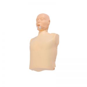
BIX/CPR100A
Advanced fully automatic electroni....
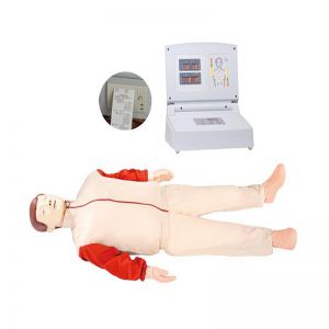
BIX/CPR480
Advanced computer half body CPR ma....
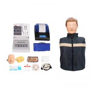
BIX/CPR260
Advanced infant head for trachea i....

BIX-J3A
Neonate Head for trachea Intubatio....
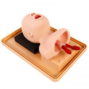
BIX-J2A
Created on:2024-06-18 | bomn
Article tag: Human skeleton model human anatomy model
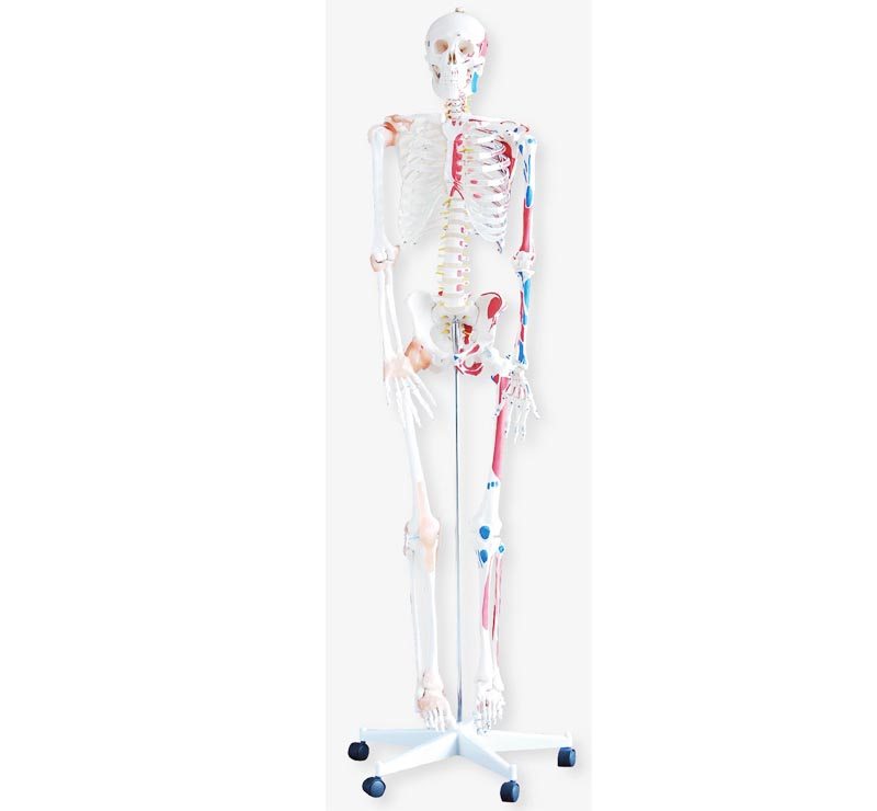
Human bone with muscle coloring and ligament model is a highly simulated medical teaching model, which integrates the structure of human bone, muscle and ligament, and clearly shows the shape, distributio...
Human bone with muscle coloring and ligament model is a highly simulated medical teaching model, which integrates the structure of human bone, muscle and ligament, and clearly shows the shape, distribution and function of these tissues in the human body through meticulous coloring treatment. This model not only provides intuitive and vivid teaching AIDS for medical education and clinical training, but also provides strong support for scientific research and academic exchange.
I. Model structure and characteristics
Bone structure: The human bone with muscle coloring and ligament model is first based on the human bone, showing the skeletal composition and shape of the whole body. These bones include the bones of the skull, spine, ribs, arms and legs, which are connected to each other and form the basic skeleton of the human body.
Muscle coloring: On the basis of bone, the model further shows the muscle tissue. By using different color processing, the model distinguishes the muscle tissue, allowing learners to clearly see the location and distribution of muscles in the human body. In general, the muscle starting point is usually colored red and the muscle stopping point is colored blue, and this color distinction helps learners better understand the function and contraction mechanism of muscles.
Ligaments Display: Ligaments are important structures that connect bones and are essential for maintaining joint stability and motor function. The ligaments are also clearly displayed in the human bone with muscle coloring and ligaments model. With specific coloring treatments, learners can clearly see where the ligaments go and where they join, thereby deepening their understanding of joint stability and movement mechanisms.
Second, function and application
Medical education: This model is an important teaching tool for medical schools, nursing schools and other medical related majors. Through observation and manipulation of models, students can intuitively understand the structure and function of human bones, muscles and ligaments, and deepen their understanding of anatomical knowledge.
Clinical training: For healthcare professionals, the model can be used for clinical skills training. By simulating surgical operations and nursing operations, medical personnel can become familiar with the human body structure and improve operational skills and safety.
Research and display: The model can also be used for research and display work in the field of scientific research. Researchers can use the model to study the structure and function of human bones, muscles and ligaments; At the same time, the model can also be used in medical exhibitions and display work to popularize medical knowledge to the public.
In short, the model of human bone with muscle coloring and ligament is a powerful and practical medical teaching model. With its highly simulated design and meticulous coloring treatment, it provides strong support and help for medical education, clinical training and scientific research fields.