


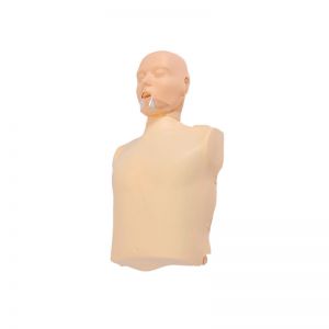
Half body CPR training manikin(Sim....
BIX/CPR100A
Advanced fully automatic electroni....
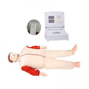
BIX/CPR480
Advanced computer half body CPR ma....
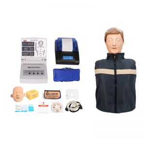
BIX/CPR260
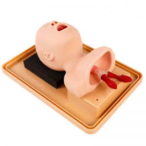
Advanced infant head for trachea i....
BIX-J3A
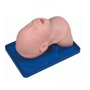
Neonate Head for trachea Intubatio....
BIX-J2A
Created on:2025-09-03 | bomn
Article tag: Hepatopancre duodenal model BIX-A1053
The hepatopancre-duodenal model is not only an anatomical display carrier, but also an important bridge between medical theory and clinical practice. It transforms complex anatomical and physiological knowledge into visible, tangible and understandable morp...
The hepatopancoduodenal model is a common tool for medical teaching and scientific research demonstration, mainly used to show the anatomical structure and functional connection among the liver, pancreas and duodenum. This model is made of simulation materials with clear color zoning, which can visually reflect the positional relationship and structural characteristics of each organ, helping learners establish a clear spatial concept in a short time.
Its design principle is to centrally display the key organs of the digestive system. For instance, the location of the liver, the direction of the gallbladder and bile ducts, the distribution of the pancreas and pancreatic ducts, as well as the shape of the duodenum and its connection with surrounding organs can all be visually observed on the model. Some models can also be disassembled for repeated operation, allowing students to understand the process by which bile and pancreatic juice enter the duodenum and master their important roles in digestion and absorption.
In medical education, this model is often used in digestive system courses, surgical operation training and pathological explanations. Through it, students can have a direct understanding of the occurrence sites and mechanisms of common diseases such as gallstones, pancreatitis and peptic ulcers. For clinicians, this model is also an effective tool for conducting health education to patients.
The hepatopancre-duodenal model is not only an anatomical display carrier, but also an important bridge between medical theory and clinical practice. It transforms complex anatomical and physiological knowledge into visible, tangible and understandable morphological manifestations, greatly enhancing the efficiency of learning and communication.