


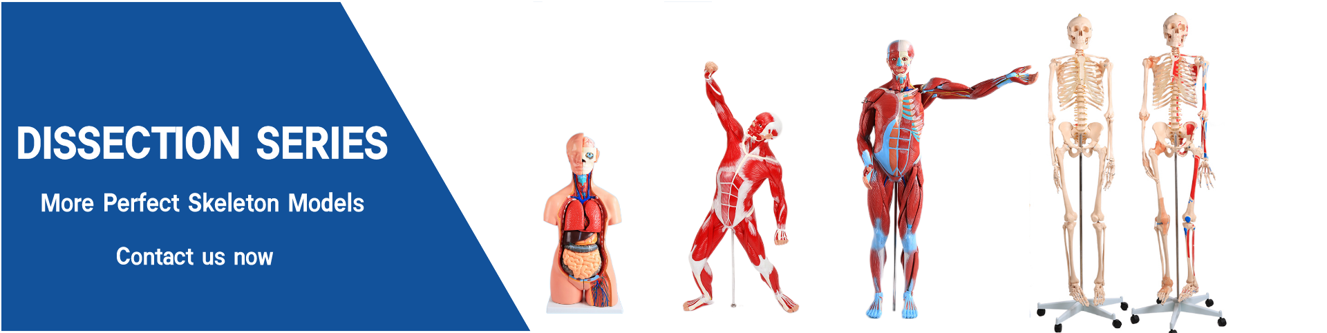
Half body CPR training manikin(Sim....
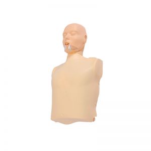
BIX/CPR100A
Advanced fully automatic electroni....
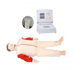
BIX/CPR480
Advanced computer half body CPR ma....
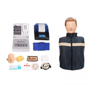
BIX/CPR260
Advanced infant head for trachea i....
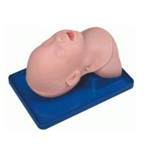
BIX-J3A
Neonate Head for trachea Intubatio....
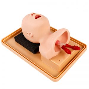
BIX-J2A
Created on:2026-11-03 | bomn
Article tag: shoulder joint model BIX-A1023 comparison shoulder motion anatomy model joint model for anatomy l

Most anatomy labs buy joint models one at a time, and the first choice is usually the shoulder or the knee. Both are in the BIX joint series — the BIX-A1023 shoulder at natural size, 17.5 × 15 × 21 cm, with a flexible artificial ligament — and both teach movement. The question is which one your syllabus needs first.
| Item | Detail |
|---|---|
| Model | BIX-A1023 |
| Category | Joint models |
| Size | Natural size, 17.5 × 15 × 21 cm |
| Movements shown | Abduction, adduction, forward tilt, backward tilt, internal and external rotation |
| Ligament | Flexible artificial ligament included |
| Setting | Anatomy, physiotherapy, orthopaedics, sports science |
| Price | Quoted per order — request the current price list |
Range-of-motion teaching. The shoulder has the widest range of any joint, and the model demonstrates it: abduction, adduction, forward and backward tilt, and both rotations. That range is the reason shoulder injuries dominate sports medicine, and the reason students must see the movements rather than read a list.
Ligament behaviour. The flexible artificial ligament lets a student feel what a restraint does at the end of range. That is a different lesson from "the ligament stabilises the joint": it shows where the restraint engages.
Clinical correlation. Frozen shoulder, rotator cuff tears and instability all map onto specific lost movements. A model that moves lets an instructor demonstrate the deficit — abduction loss versus external rotation loss — in a way a chart cannot.
A knee model in the same series (BIX-A1025) is the better first purchase if your programme is orthopaedic or sports-focused, because knee mechanics — the screw-home mechanism, meniscal movement, collateral and cruciate restraint — are the content most often examined. The shoulder wins when the curriculum is general anatomy or physiotherapy, where upper-limb range dominates.
The decision rule is simple: buy the joint whose movements appear most often in your exam questions.
Anatomy teaching research reports a measurable advantage for three-dimensional models over flat images in spatial understanding [1]. Motion adds a second dimension: students see that a joint is a constraint system, not a hinge with a name. The learning gain shows up in viva questions — students who have moved a model describe the lost movement in a lesion question instead of reciting muscle names.
The shoulder is the clearest example of that trade-off in the body. It gives up bony congruence for range, and depends on capsule, labrum and the rotator cuff for restraint [3]. A model whose artificial ligament tightens only at end range reproduces exactly where those restraints fail — and because the pattern is learned by repetition rather than by a single demonstration [2], the 25-minute session below ends with retrieval, not with a summary.
Q1. Which movements does the BIX-A1023 show? Abduction, adduction, forward tilt, backward tilt, and internal and external rotation.
Q2. Is it life-size? Yes — natural size, 17.5 × 15 × 21 cm.
Q3. Does it include ligaments? Yes, a flexible artificial ligament is part of the model.
Q4. Is it suitable for physiotherapy teaching? Yes — range-of-motion demonstration is its primary use.
Q5. What is the minimum order quantity? MOQ is flexible and quoted per order. Email for the price list and volume tiers.
Q6. How do we request pricing? Email cprwell@adaanatomy.com with quantity, city and course type for the price list, packing data and lead time.
For the specification sheet, ligament replacement and a lab quotation: cprwell@adaanatomy.com. Production follows ISO 45001 management practice; CE documentation is available for EU procurement.