


Half body CPR training manikin(Sim....
BIX/CPR100A
Advanced fully automatic electroni....
BIX/CPR480
Advanced computer half body CPR ma....
BIX/CPR260
Advanced infant head for trachea i....
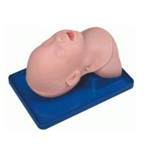
BIX-J3A
Neonate Head for trachea Intubatio....
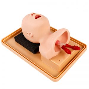
BIX-J2A
Created on:2026-11-19 | bomn
Three-part heart model enlarged 4x (25x23x23 cm, PVC) for classroom teaching of chambers, valves and great vessels.
...The BIX-A1067 is a three-part heart model enlarged four times, measuring 25 × 23 × 23 cm and made of PVC. It is built for classroom demonstration of external shape, internal chambers and the great vessels — the pathway question students most often fail to answer from a textbook diagram.
Quick answer. The A1067 is a lecture-room model, not a dissection trainer. At 4× scale, a class of 30 can see the valve plane and vessel origins from the back row. It does not show coronary circulation in detail or the conduction system.
| Item | Specification | Classroom effect |
|---|---|---|
| Scale | 4× life size | Visible from several metres away |
| Parts | 3, separable | Opens along the chamber plane |
| Size | 25 × 23 × 23 cm | Fits a demo bench, not a student desk |
| Material | PVC | Wipeable, durable, no odour |
| Packing | 8 per box, 58 × 45 × 50 cm, 16 kg | Freight is per box, not per unit |
| Contents | Heart with great vessels | No removable organ set |
A life-size heart is about the size of a fist. In a 30-student lecture theatre, that is a small object at 8 metres. The 4× model makes the pulmonary trunk, aorta, superior vena cava and valve plane legible without a camera, which changes how much time is spent narrating versus showing.
The trade-off is that scale distorts proportion perception. Students who learn only on an enlarged model tend to underestimate how compact the mediastinum is. Use the 4× model for structure and a life-size or digital model for spatial relations in the chest.
The model separates to expose the chamber anatomy, so the instructor can open it mid-sentence rather than passing around a closed cast:
A useful sequence is external → open → trace. Students trace a drop of blood from vena cava to pulmonary artery, then from pulmonary vein to aorta, saying each chamber and valve aloud. The model cannot show flow, so the narration is what carries the physiology.
Q1. How large is the BIX-A1067? Four times life size, 25 × 23 × 23 cm.
Q2. How many parts does it have? Three separable parts showing external surface, chambers and great vessels.
Q3. What material is it made of? PVC.
Q4. Does it show the conduction system? No. The nodes and conduction pathways are not modelled.
Q5. Is it suitable for medical students or school biology? Both — the product page specifies general secondary and specialised schools. The depth of detail suits introductory anatomy.
Q6. How is it packed? Eight units per carton, 58 × 45 × 50 cm, 16 kg.
Related on this site: medical first aid model collection · all articles
Contact. For carton pricing, spare parts or a teaching-set quote, email cprwell@adaanatomy.com.