


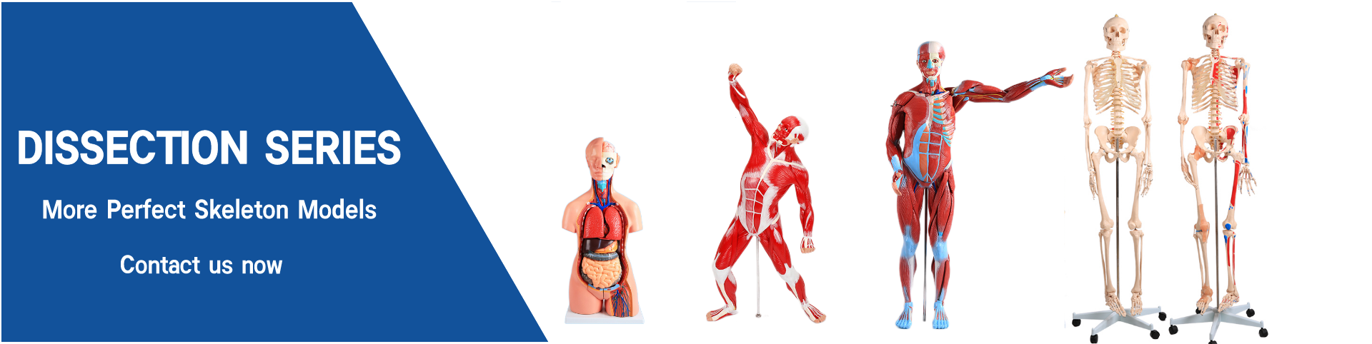
Half body CPR training manikin(Sim....
BIX/CPR100A
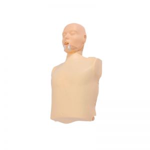
Advanced fully automatic electroni....
BIX/CPR480
Advanced computer half body CPR ma....

BIX/CPR260
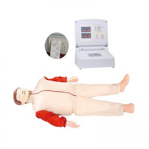
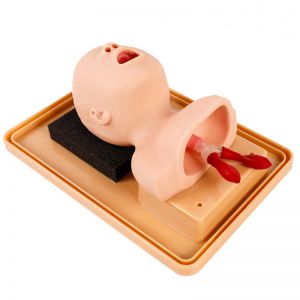
Advanced infant head for trachea i....
BIX-J3A
Neonate Head for trachea Intubatio....
BIX-J2A
Created on:2025-09-09 | bomn
Article tag: Middle head section model BIX-A1065
The mid-head section model is a teaching aid commonly used in anatomy instruction and clinical training. It involves cutting the head along a sagittal or coronal direction to display the internal structures of the skull, brain tissue, nasal cavity, pharynx,...
The mid-head section model is a teaching aid commonly used in anatomy instruction and clinical training. It involves cutting the head along a sagittal or coronal direction to display the internal structures of the skull, brain tissue, nasal cavity, pharynx, etc., helping learners gain a deeper understanding of the spatial relationships within the skull. During the operation process, the following steps can generally be followed
First, check the integrity and structure of the model to ensure that the cross-section is clear and all components are complete. Place the model on a stable stand or platform to prevent it from sliding or toppling over during operation.
Secondly, observe in accordance with the teaching objectives. The user should start from the overall perspective, identify the cross-sectional positions of the skull and brain tissue, and then successively search for key structures such as the ventricular system, nasal cavity, throat and related blood vessels and nerves. Some models are marked with colors or numbered. Learners can refer to the diagrams or instruction manuals for study to enhance their memory.
Secondly, if the model has detachable parts, they can be gradually disassembled to simulate the clinical or anatomical observation process. When disassembling and resetting, handle with care to avoid damaging the interface due to excessive force.
Finally, after the operation is completed, the model should be cleaned, organized and properly stored to keep the cut surface clear and the durability of the components.
Overall, the mid-head section model, through systematic operation steps, not only helps students master the spatial relationship of the internal anatomical structure of the skull, but also enhances their cognitive ability in clinical diagnosis and surgical planning.