


Half body CPR training manikin(Sim....
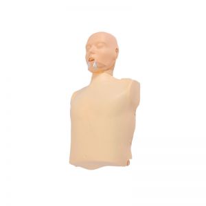
BIX/CPR100A
Advanced fully automatic electroni....
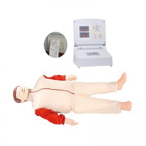
BIX/CPR480
Advanced computer half body CPR ma....
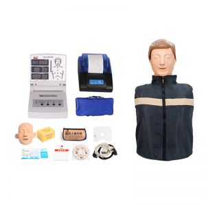
BIX/CPR260
Advanced infant head for trachea i....
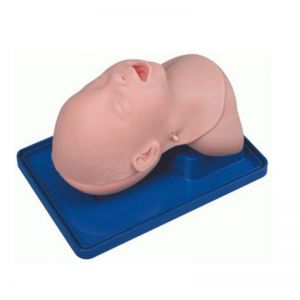
BIX-J3A
Neonate Head for trachea Intubatio....
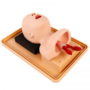
BIX-J2A
Created on:2026-09-03 | bomn
Article tag: Cervical vertebra model BIX-A1021 A1021
A training guide for the BIX-A1021 cervical model — occipital bone and seven cervical vertebrae with intervertebral discs, cervical arteries, and nerve structures on a plastic base, for neck anatomy teaching in chiropractic, physiotherapy, and medical pro...
Model | BIX-A1021 — Cervical Model with Carotid Artery, Occipital Bone, Disc & Nerve |
Summary | Cervical spine model: occipital bone, 7 cervical vertebrae with discs, nerves and arteries, on plastic base. For neck anatomy teaching. (135 chars) |
Price | On request |
Included Anatomy | Occipital bone; 7 cervical vertebrae (C1–C7); intervertebral discs; cervical nerve and artery structures |
Mounting | Plastic base |
Category | Skeleton Models — anatomy teaching |
Audience | Chiropractic, physiotherapy, medical, nursing, and sports-medicine programs |
Educational-use note: anatomy teaching aid — educational equipment, not a medical device or pharmaceutical product, and contains no medicinal ingredients. Confirm the exact arterial/nerve display (per official specification: cervical vertebral artery with carotid structures in the product title) with the supplier.
The cervical spine is the most complex and most vulnerable region of the vertebral column: seven vertebrae (C1–C7) articulate with the occipital bone, carry the weight of the head, protect the spinal cord, and transmit the vertebral arteries through their transverse foramina — while intervertebral discs and emerging nerve roots occupy the tight spaces where pathology concentrates.
Its clinical burden is enormous. Neck pain affected an estimated 203 million people globally in 2020, remains a leading contributor to disability worldwide, and case numbers are projected to keep rising with population ageing (GBD Neck Pain Collaborators, 2024).
Yet the cervical region is also where anatomy is hardest to teach from a single generic skeleton:
● The occipito-atlanto-axial complex (C0–C1–C2) is functionally distinct from the lower cervical spine — generic models cannot show it.
● Discs and nerve roots are invisible on a bare skeleton.
● The arterial relations that make neck handling and manipulation a high-stakes clinical skill are entirely absent.
A dedicated cervical model makes this crowded anatomy visible, ordered, and repeatable — which is precisely what novices need before they touch a real patient.
Evidence | Finding | Implication |
Fleming et al., 2020 | Randomized-trial meta-analysis: three-dimensional physical models were associated with significantly higher anatomy exam scores in medical students than 2-D methods (P<0.0001) (J Am Coll Radiol 2020) | Physical models measurably outperform flat teaching for novices |
Cook et al., 2011 | Technology-enhanced instruction produces large learning gains — effect sizes 1.09–1.20 across 35,226 learners (JAMA 2011) | Model-based teaching is among the most effective known methods |
GBD Neck Pain Collaborators, 2024 | 203 million people affected by neck pain in 2020; major disability burden globally (Lancet Rheumatol 2024) | Cervical anatomy knowledge underpins care for a top disability cause |
The teaching logic is direct: structures that students can see, touch, and rotate on a model are structures they can identify in a clinic. For cervical spine assessment — where a few millimetres of anatomy separate safe from unsafe practice — that visibility is not a luxury.
Teaching Module | Anatomy Shown | Clinical Context |
Occipito-cervical junction | Occipital bone + C1/C2 relationships | Head-neck transition; the junction assessed in every neck examination |
Vertebral column segment | C1–C7 with discs | Segmental anatomy; disc-related cervical pathology |
Nerve structures | Cervical nerve display | Radicular pain patterns in physiotherapy and chiropractic |
Arterial relations | Cervical arteries (per official spec) | Why vascular awareness matters in neck assessment and handling |
Exact display of arterial vs nerve structures varies by production run — confirm the specification sheet with your supplier.
1. Identify the occipital bone and each cervical vertebra (C1–C7) on the model.
2. Trace the disc spaces and the nerve-root levels.
3. Relate each landmark to a palpation site used in clinical examination.
1. Demonstrate C0–C1–C2 movement relationships on the model.
2. Discuss why this region is assessed separately in neck examination.
1. Show where discs bulge, where nerve roots exit, and the arterial course.
2. Link each relationship to a clinical scenario (e.g., radicular pain, vascular caution).
Refresher note: anatomy recognition, like other clinical knowledge, decays without reinforcement — re-use the model in every spinal-palpation refresher block.
Skill | Standard |
Vertebra identification | Names C1–C7 and occipital bone correctly |
Disc level location | Locates disc spaces between named vertebrae |
Nerve-root mapping | States the nerve level at each exit point |
Clinical translation | Links each structure to a palpation/assessment step |
Item | Frequency | Notes |
Surface cleaning | After each class | Wipe with mild soap and damp cloth |
Base/seat | Check monthly | Verify the plastic base is stable |
Painted landmarks | Per manual | Confirm color-coding remains legible |
Storage | Always | Dry, dust-free; follow manual storage |
Q1: What exactly is included on the A1021? A: Per the official specification: the occipital bone and seven cervical vertebrae with a cervical artery, displayed on a plastic base. The product title also references disc and nerve structures — request the specification sheet to confirm the exact painted display of your production unit.
Q2: What is the difference between A1021 and full skeleton models like A1004? A: A1021 is a region-specific cervical model — it shows the occipito-cervical junction, discs, nerves, and arteries in detail that a whole skeleton (A1004, 85 cm) cannot provide. Programs teaching neck assessment typically use both: the full skeleton for whole-body context, the A1021 for cervical detail.
Q3: Which programs benefit most from this model? A: Chiropractic and physiotherapy (cervical assessment and manipulation training), medical anatomy courses, nursing, sports medicine, and any curriculum covering neck pain — a condition affecting 203 million people globally (GBD Neck Pain Collaborators, 2024).
Q4: Is it useful for teaching cervical pathology? A: Yes for structure-based teaching: disc spaces and nerve-root levels are the anatomical reference for understanding cervical radiculopathy and disc pathology. It is a teaching model, not a pathology demonstration model — confirm if a pathology-specific version is needed.
Q5: Is this a medical device? A: No — it is educational equipment for anatomy teaching, contains no medicinal ingredients, and is not a pharmaceutical product or clinical device.
Q6: What is the MOQ and delivery time? A: MOQ is 1 unit. Air freight: 7–10 business days; sea freight: 30–45 days. Email cprwell@adaanatomy.com for a quote.
Global, Regional, and National Burden of Neck Pain, 1990–2020, and Projections to 2050 — GBD Neck Pain Collaborators (2024), Lancet Rheumatol 6(3):e142–e155
Effectiveness of Three-Dimensionally Printed Models in Anatomy Education for Medical Students and Resident Physicians: Systematic Review and Meta-Analysis — Fleming et al. (2020), J Am Coll Radiol 17(10):1220–1229
Technology-Enhanced Simulation for Health Professions Education: A Systematic Review and Meta-analysis — Cook et al. (2011), JAMA 306(9):978–988