



Half body CPR training manikin(Sim....
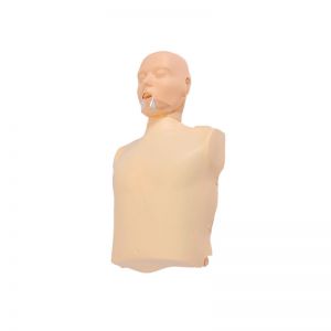
BIX/CPR100A
Advanced fully automatic electroni....
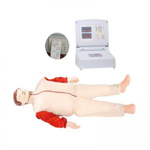
BIX/CPR480
Advanced computer half body CPR ma....

BIX/CPR260
Advanced infant head for trachea i....
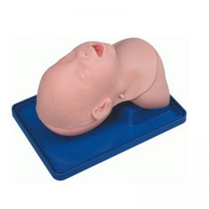
BIX-J3A
Neonate Head for trachea Intubatio....

BIX-J2A
Created on:2026-09-14 | bomn
Article tag: Human Torso Coronal Sectional Anatomical Model BIX-A3009 A3009
BIX-A3009 human torso coronal sectional anatomical model — 8 parts showing internal organs in coronal (frontal) section, for sectional anatomy, radiology and medical training. Model-building raised CT examination scores (Oh et al., 2009); 3D beat 2D cross...
Model | BIX-A3009 — Human Torso Coronal Sectional Anatomical Model (8-Part Frontal Section Torso) |
Summary | Human torso coronal sectional anatomical model: an 8-part set showing internal organs in coronal (frontal) section for sectional anatomy teaching. |
Configuration | A set of 8 parts (official specification) |
Section Plane | Coronal (frontal) — organs displayed as in a frontal cut |
Stated Use | Sectional anatomy teaching, radiology and medical training |
Companion Models | A3004/A3005 sagittal-slice torsos add the sagittal plane |
Certification | ISO & CE (as stated on the product page) |
Listed Price | US $1,478.06 per set (confirm by quotation) |
Audience | Medical schools, radiology teaching, nursing faculties |
Educational-use note: anatomical teaching model — educational equipment, not a medical device. Request the spec sheet by email for the part list and options.
Students meet anatomy in one plane far more often than the others. Axial (transverse) sections dominate imaging teaching, textbooks and model kits — yet clinicians read coronal views daily: frontal reconstructions, chest radiographs, and the frontal relationships that explain organ displacement. A student who thinks only in axial slices recognises structures on a CT scroll but loses orientation when the plane changes.
That asymmetry is documented. In a study that added clay modelling to anatomy courses, students built models, cut them, and compared the cut surfaces with CT and MR images to see how two-dimensional cross-sections arise from three-dimensional structure. The clay group's average CT examination score was higher than that of the group that did not use models, the approach proving most effective where complex cross-sectional understanding is required (Oh et al., 2009).
The A3009 puts that principle into a durable classroom object: a human torso in coronal section as a set of 8 parts, so organs normally met as flat images can be handled, separated and reassembled in the plane radiographs use.
The same evidence base warns against expecting a model to teach by itself. Physical anatomy models are typically low fidelity — a subset of structures, without accurate surface detail — but they correspond strongly in spatial relationships, which makes them memory aids that reduce cognitive overload and stimulate problem solving. Model use should be accompanied by explanation to establish the correspondence between model and body (Chan & Cheng, 2011).
Evidence | Finding | Relevance to A3009 |
Oh et al., 2009 | Model-building group scored higher on the CT examination than a non-model group; best for complex content | Physical sectioning teaches cross-sectional correlation |
Estevez et al., 2010 | RCT: physical model group scored significantly higher, gain from 3D questions (P = 0.02); 84% wanted it repeated | Plane-specific gains, not general enthusiasm |
Ruisoto et al., 2012 | 80 participants: 3D gave higher accuracy and confidence and lower response time than 2D cross-sections, most on difficult images | 3D form beats flat cross-sections |
Wainman et al., 2018 | Physical models 67–69% vs 41% for screen learning; 74% vs 43% in transfer | Physical handling outperforms on-screen study |
Chan & Cheng, 2011 | Low-fidelity models work through spatial correspondence; need explanation alongside | How to use the A3009 correctly |
Khalil et al., 2008 | Computer-based cross-section strategies gave no significant benefit and raised cognitive load | Screen-only cross-sections are insufficient |
The coronal configuration makes the translation step explicit: identify a structure on the cut surface, then find it on a frontal image. That translation — not the naming — is the skill radiology teaching aims at, and the step most students skip.
With 8 parts, the model supports a removal-and-rebuild sequence in which coronal relationships are reconstructed, not memorised. Reassembly tests recall in reverse and exposes which relationships were never learned.
Sectional anatomy needs sagittal and coronal as well as axial. Pairing the A3009 (coronal) with the A3004/A3005 sagittal slices lets one class study both non-axial planes on consistent physical models.
A frontal section is the natural view for midline structures and for showing how one side relates to the other — useful when explaining why lesions displace rather than compress.
Because low-fidelity models need explanation to work (Chan & Cheng, 2011), the A3009 supports a lesson on what the model shows, what it omits, and how to check it against real imaging.
Model | Format | Parts | Best for |
A3009 | Coronal (frontal) section torso | 8 | Sectional anatomy, radiology correlation |
A3004 | Male sagittal slice | 10 | Sagittal plane, male anatomy |
A3005 | Female sagittal slice | 10 | Sagittal plane, female anatomy |
A1043 | 85 cm both-sex torso | 40 | Full-size demonstration, organ detail |
A1046 | 45 cm both-sex torso | 23 | Comparative organ study |
A1047 | 42 cm asexual torso | 18 | Student stations |
Buying logic: choose the A3009 for plane-based reasoning — reading coronal images, translating between model and radiograph. Add A3004/A3005 to complete the non-axial planes; choose A1043/A1047 when the objective is organ detail.
Station | Time | Activity |
A. Orientation | 10 min | Establish the coronal plane; contrast with axial and sagittal |
B. Identify | 15 min | Name structures on the intact cut surface |
C. Disassemble | 20 min | Remove the 8 parts in order; one relationship per part |
D. Image match | 20 min | Match structures to a frontal image; state what is omitted |
E. Rebuild | 15 min | Reassemble unlabelled; peer-check |
● Coronal plane distinguished from axial and sagittal
● Required structures named on the intact model
● Parts removed and reseated in order without forcing
● Each structure located on a frontal image
● Two model limitations stated and checked against imaging
● Torso reassembled with all 8 parts correctly seated
Item | Frequency | Notes |
Cut surfaces and parts | Each session | Mild disinfectant; no solvents |
Fittings | Monthly | Check seating points |
Storage | Daily | Parts in position; away from sun |
Q1: What is the BIX-A3009? A: A human torso coronal sectional anatomical model supplied as 8 parts, showing internal organs in coronal (frontal) section, for sectional anatomy, radiology and medical training.
Q2: What is a coronal section, and why does it matter? A: A coronal (frontal) cut divides the body front-to-back — the plane used for chest radiographs and frontal reconstructions. Teaching over-emphasises axial sections, so students struggle when the plane changes; this model targets that gap.
Q3: How many parts are included? A: Eight parts per set (official specification). Request the spec sheet at cprwell@adaanatomy.com. for the part list.
Q4: Does it include a sagittal model too? A: No. Add the A3004 (male) and A3005 (female) sagittal-slice torsos to cover the sagittal plane alongside the coronal A3009.
Q5: What is the price and MOQ? A: Listed at US $1,478.06 per set; MOQ 1 set. Final pricing depends on configuration and volume — email cprwell@adaanatomy.com. for the current quotation.
Q6: What certifications and shipping terms apply? A: ISO & CE as stated on the product page; air freight 7–10 days, sea freight 30–45 days.
Clay Models and Cross-Sectional Anatomy (Oh et al., 2009)
Physical Brain Models in Neuroanatomy (Estevez et al., 2010)
3D vs 2D Cross-Sections in Imaging (Ruisoto et al., 2012)
Physical Models vs Screen Learning (Wainman et al., 2018)
Educational Value of Anatomy Models (Chan & Cheng, 2011)
Cross-Section Strategies and Cognitive Load (Khalil et al., 2008)